Mechanical properties of chordotonal organs
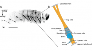
Most if not all animals require reliable mechanosensation for various biological processes. Vertebrates and invertebrates have evolved specialized sensory devices and strategies to manage this immense challenge. For most mechanosensation, however, the underlying physics of living materials (cells, tissues, organs) has remained elusive. These constitutive material properties, including elasticity and isometric tension, imply fundamental gaps in our knowledge of how sensory systems work. The fruit fly Drosophila melanogaster possesses specialized organs, chordotonal organs (ChOs), to “hear” external sound, feel airflow, and keep track of body motions (propiosensing). Combining mechanical micromanipulation with the help of calibrated glass needles and electrophysiological recordings from the sensory neurons, I have established an experimental platform to measure the material properties of Drosophila larval ChOs and characterize their neuronal response, adaptation, and saturation. I propose to use the ChO as an in vivo model to study the roles of mechanical players such as extracellular matrix (ECM), molecular motors, and the cytoskeleton, in adjusting cell mechanics and sensory transduction. I aim at exploring the molecular mechanisms involved in the regulation and encoding of mechanical forces by primary mechanoreceptor neurons.
